
Molecular Docking: From In-Silico Mechanics to Industrial R&D Pipeline
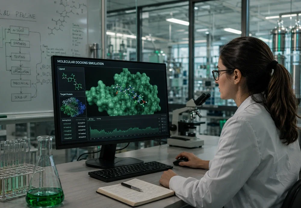
Molecular Docking: From In-Silico Mechanics to Industrial R&D Pipeline

Chief Product Officer at ChemCopilot. Former VP of Product at Noble.AI, Co-founder/CTO at Nanostellar (Quantum Simulation & Catalysts), and NASA/Harvard ACIS Software Team Leader. Over 25 years of expertise pioneering quantum mechanical simulations, molecular docking workflows, and enterprise SaaS.
1. What is Molecular Docking?
At its core, molecular docking models the atomic-level interaction between a small molecule (ligand) and a macromolecular target (protein, enzyme, or nanomaterial). It resolves three critical parameters:
- Binding Affinity ($\Delta G$): Quantifies interaction strength in $\text{kcal/mol}$ or $\text{kJ/mol}$ using empirical or physics-based scoring functions.
- Pose Prediction: Determines the lowest-energy 3D spatial orientation of the ligand within the target binding site.
- Binding Pocket Mapping: Pinpoints key hydrogen bonds, hydrophobic pockets, and electrostatic contacts driving binding stability.
2. Required Software Comparison
Choosing the right docking suite depends on your research scope, required throughput, and technical expertise:
| Software | Type | Best For | Learning Curve |
|---|---|---|---|
| ChemCopilot | Free Tier / Enterprise | Automated Docking, Substitution & Sustainability PLM | Very Low (Zero-Code) |
| AutoDock Vina | Open-Source (Free) | Beginners, Academic Scripting & Virtual Screening | Low |
| Schrödinger Suite (Glide) | Commercial (Paid) | Industry Pharma Professionals & FEP+ Simulations | High |
| UCSF Chimera / PyMOL | Free / Freemium | Visualizing Binding Poses & Surface Maps | Medium |
| GROMACS / NAMD | Open-Source (Free) | Advanced Molecular Dynamics (MD) Pose Refinement | Very High |
3. Step-by-Step Docking Tutorial (AutoDock Vina)
Step 1: Molecule Preparation
- Fetch receptor crystal structure from the RCSB Protein Data Bank (e.g., PDB ID:
6LU7). - Remove crystallographic water molecules, assign Gasteiger charges, and add polar hydrogens using AutoDockTools or PyMOL.
- Save receptor as
protein.pdbqtand ligand asligand.pdbqt.
Step 2: Define Grid Search Space
Enclose the target active site within a defined 3D bounding box coordinates:
# Search Space Envelope (grid_box.conf)
center_x = 15.0
center_y = 12.5
center_z = 10.0
size_x = 25.0
size_y = 25.0
size_z = 25.0
exhaustiveness = 8
Step 3: Execute Docking Simulation
# Run AutoDock Vina via CLI
vina --receptor protein.pdbqt \
     --ligand ligand.pdbqt \
     --center_x 15.0 --center_y 12.5 --center_z 10.0 \
     --size_x 25.0 --size_y 25.0 --size_z 25.0 \
     --out output_poses.pdbqt \
     --log docking_log.txt
Step 4: Pose Evaluation & Visualization
Evaluate binding score output ($\text{kcal/mol}$). More negative values indicate higher thermodynamic stability (e.g., $-8.5\text{ kcal/mol}$ is stronger than $-5.2\text{ kcal/mol}$). Load into PyMOL to verify polar hydrogen contacts:
# PyMOL Command Line
load protein.pdb
load output_poses.pdbqt
show sticks, ligand
distance h_bonds, protein, ligand, 3.2, mode=2
4. Common Pitfalls & Troubleshooting
| Observed Issue | Root Cause | Recommended Expert Solution |
|---|---|---|
| Unrealistic Binding Pose | Grid box too small or offset from true pocket | Expand box boundaries to $30\text{Å}^3$ or use blind docking algorithms. |
| Inaccurate Affinity Score | Incorrect protonation states at physiological pH (7.4) | Recalculate pKa using OpenEye or ChemCopilot automated state assigners. |
| Simulation Crash | Missing atom types or excessive flexible bonds (>32) | Freeze non-critical ring torsions or switch to flexible-receptor models. |
